Endoscopic Zygoma Reduction
This surgery reduces the lateral cheekbone that makes the face appear wide, through a small incision within the temporal hairline rather than inside the mouth.
- Where are the incisions?
- 2–3 cm within the temporal hairline and 8 mm at the sideburn. No incision is made inside the mouth.
- How is the bone reduced?
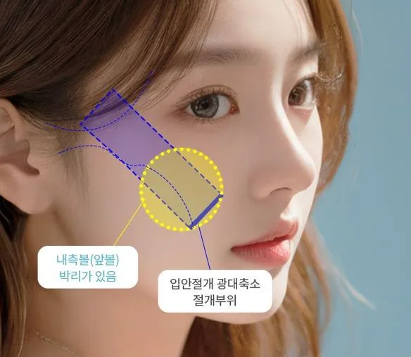
- The posterior part of the lateral cheekbone is cut obliquely and moved inward while part of the anterior bone connection is preserved. The oblique cut surfaces interlock and hold the bone in position without plates.
- Cheek sagging
- Leaving the anterior cheek tissue attached to the bone reduces the likelihood of cheek sagging.
- Anesthesia and recovery
- The procedure uses sedation rather than general anesthesia. UVOM Plastic Surgery advises that same-day discharge and eating on the day of surgery are possible.
How the Approaches Differ
Conventional approach
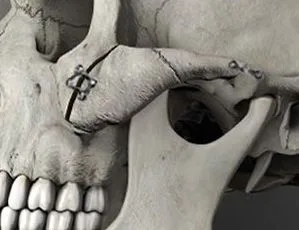
Zygoma reduction is usually performed through an incision inside the mouth above the upper gum. The inner cheek tissue is separated from the bone to expose the cheekbone, which is then cut, moved inward and secured with small plates and screws. General anesthesia is usually used.
Limitations
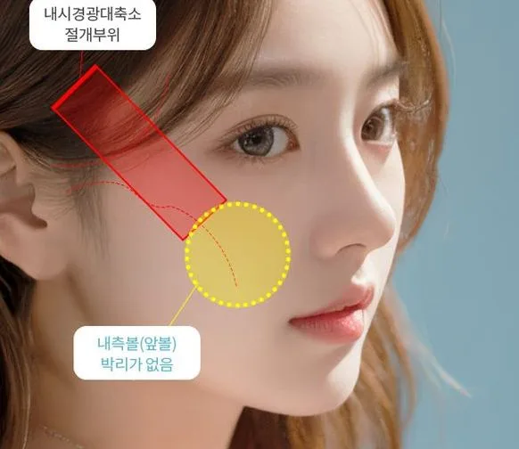
Exposing the bone requires extensive separation of the cheek tissue from it. The tissues anchoring the cheek to the bone are also detached. The bone becomes smaller while the overlying soft tissue remains, so the anterior cheek may bulge or sag downward.
An intraoral wound can make eating and brushing teeth uncomfortable for a time and requires infection management. The fixation plates and screws remain in the body.
Dr. Kang’s approach
No incision is made inside the mouth. Small incisions are made within the temporal hairline and at the sideburn. An endoscope, a thin camera, is inserted so surgery can be performed while viewing a monitor.
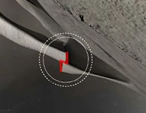
The long, slender lateral cheekbone running from in front of the ear to the cheekbone contributes to the appearance of facial width. Its posterior portion is completely cut at an oblique angle, while part of the anterior connection is left intact. When the bone is then pushed inward, the oblique cut surfaces interlock and remain in position. Seen from above, they form a Z-shaped interlock.
Benefits and considerations
Leaving the anterior cheek tissue attached to the bone reduces the likelihood of cheek sagging. Without an intraoral wound, eating is more comfortable, and no plates or screws are used. The incisions lie within the hair, making scars less noticeable.
This approach is not suitable for every face. When the anterior cheekbone is very prominent or substantial movement is required, an intraoral approach with plate fixation is safer. Reduced sensation, asymmetry and impaired bone healing can occur, so preoperative CT is used to assess suitability.
Glossary
- Lateral cheekbone (zygomatic arch)
- The long, slender bone running from in front of the ear to the cheekbone. It determines facial width when viewed from the front.
- Anterior cheekbone (zygomatic body)
- The fuller bone at the front of the cheek below the eye. It contributes to projection in profile.
- Osteotomy
- Cutting a bone.
- Endoscope
- An instrument with a camera at the end of a thin tube. It is inserted through a small incision to provide a magnified view of the inside on a monitor during surgery.
- J-SAW
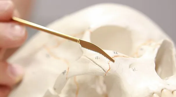
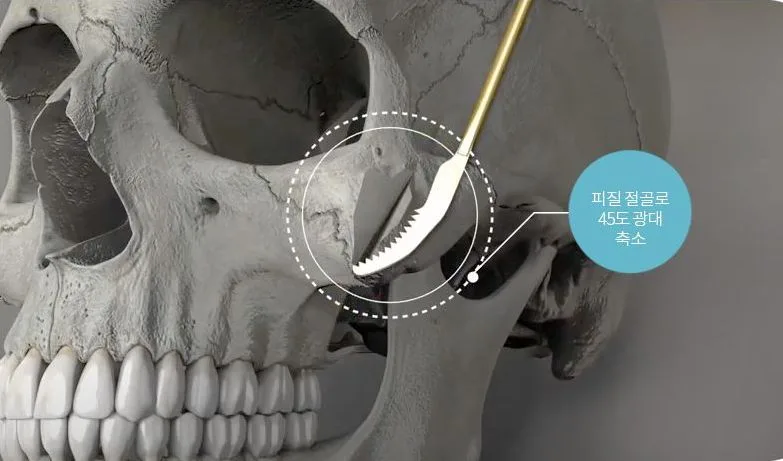
- A specialized saw with a J-shaped tip designed to cut the lateral cheekbone through a narrow passage.
Cheekbone Width and the Zygomatic Arch
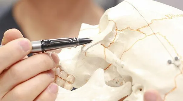
Facial width is usually associated with the lateral cheekbones. The oblique junction between the lateral and anterior cheekbone is called the 45-degree cheekbone region. Its volume can be reduced by shaving a thin layer from the outer bone surface.
This operation does not shave down the anterior cheekbone. It preserves anterior cheek projection while moving the lateral cheekbone inward, creating a smoother transition between the two.
Comparison with the Intraoral Approach
Intraoral zygoma reduction uses an incision above the upper gum and separates the cheek tissue from the bone to expose it. The tissues anchoring the cheek to the bone are also detached. As the bone becomes smaller while the overlying tissue remains, the anterior cheek may bulge or sag.
| Feature | Endoscopic zygoma reduction | Intraoral zygoma reduction |
|---|---|---|
| Incision sites | 2–3 cm in the temporal scalp; 8 mm at the sideburn | Oral mucosa; sideburn if needed |
| Anterior cheek tissue | Not detached from the bone | Detached from the bone, including the tissues anchoring the cheek |
| Fixation | Oblique cut surfaces interlock in a Z shape | Titanium plates and screws |
| Anterior cheekbone | Projection is preserved; the 45-degree region is adjusted by contouring the outer bone surface | An L- or I-shaped osteotomy allows movement of the anterior cheekbone as well |
| Anesthesia | Sedation | Usually general anesthesia |
| Suitable cases | Those seeking reduction of the lateral cheekbone while preserving the anterior cheekbone | Marked prominence of both the 45-degree cheekbone region and lateral cheekbone |
Surgical Steps
The lateral cheekbone is reduced through an incision within the temporal hairline, and the cut surfaces form a Z-shaped interlock for stability. The procedure has five steps.
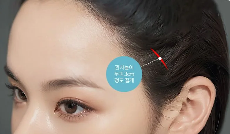
Incision within the temporal hairline
After planning the surgery, an incision of approximately 3 cm is made within the temporal hairline to make the scar less noticeable.
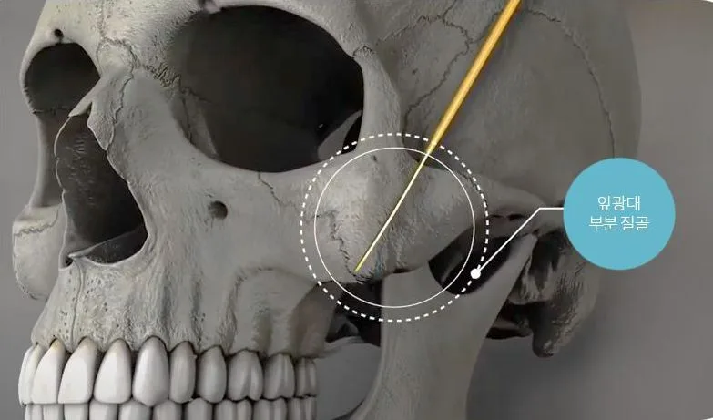
Partial anterior cheekbone osteotomy
The specialized J-SAW cuts the 45-degree anterior cheekbone region. The lower end is left intact to maintain a bony connection.
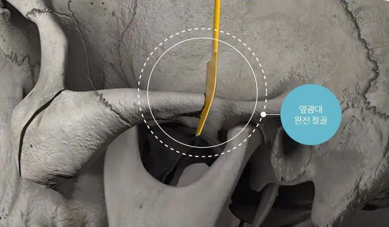
Complete lateral cheekbone osteotomy
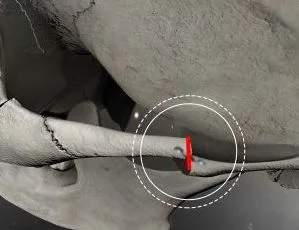
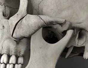
The lateral cheekbone is completely cut at an oblique 45-degree angle rather than vertically.
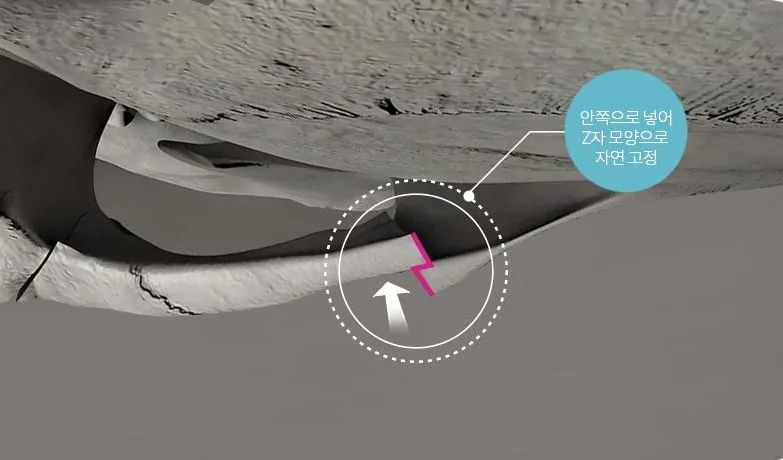
Inward rotation of the cheekbone
The cut lateral cheekbone is pushed and rotated inward. The cut surfaces interlock in a Z shape, holding the bone in position without fixation pins.
Contouring the 45-degree cheekbone region
If necessary, the outer bone surface of the 45-degree anterior cheekbone region is contoured to reduce its size.
How the Bone Is Stabilized
Intraoral zygoma reduction completely separates the cheekbone segment, moves it inward and secures it with plates. Endoscopic zygoma reduction preserves the inner connection of the anterior bone while cutting the posterior portion, allowing it to rotate inward like a closing door. The preserved anterior connection means separate fixation is not required.
Cutting the posterior portion obliquely at 45 degrees allows the cut end to slip beneath the remaining bone and interlock. Seen from above, this forms a Z shape.
Intraoral zygoma reduction
Endoscopic zygoma reduction
Conference Presentations and Supporting Papers
Dr. Chang Min Kang presented infracture using an impactor at the international congress of the Korean Society of Plastic and Reconstructive Surgeons in 2022. Related publications and textbooks are listed below.
- 2022
- Endoscopic Zygoma Reduction: Infracture Using an ImpactorPRS KOREA 2022, international congress of the Korean Society of Plastic and Reconstructive Surgeons · Chang Min Kang
- 2021
- Endoscopically assisted malarplasty: L-rotation techniqueJ Stomatol Oral Maxillofac Surg. 2021;122(3):229-234 · 261 cases · Jung Su Lee
- 2013
- Revision malarplasty using the EZ methodArch Aesthetic Plast Surg. 2013;19(2):95-100 · 11 revision cases · Jung Su Lee
- 2003
- Endoscopically assisted malarplasty: one incision and two dissection planesPlast Reconstr Surg. 2003;111(1):461-7 · 32 cases · Jung Su Lee
Indications for Intraoral Zygoma Reduction
When both the 45-degree region and lateral cheekbone are very prominent, a broad L-shaped osteotomy extends from the maxilla to the cheekbone, allowing inward movement and fixation with titanium plates. When the 45-degree region is small but the lateral cheekbone is prominent, an I-shaped osteotomy is used. Different techniques are used on each side when there is asymmetry.
To reduce cheek sagging with an intraoral approach, cheek tissue is detached only as much as necessary, excess inner cheek fat is reduced appropriately, and firm plate fixation supports bone healing.
Who May Be a Candidate
- Those seeking a gently curved cheekbone contour
- Those seeking lateral cheekbone reduction while preserving anterior cheek volume
- Those whose prominent cheekbones on both sides make the face appear wide
- Those with cheekbone asymmetry
- Those with prominent lateral cheekbones

Surgery and Consultation
Dr. Chang Min Kang, a specialist in plastic surgery in Korea, provides care. About the doctor
Risks and Limitations
Zygoma reduction can cause reduced sensation, cheek sagging, nonunion or malunion, asymmetry, limited mouth opening, bleeding and infection; facial nerve injury has also been reported rarely. Small incisions do not eliminate risk. The location of prominence, direction of movement, bone contact and stability, and soft-tissue support are assessed together. When the anterior zygomatic body is very prominent or a bone segment requires substantial movement and rigid fixation, an intraoral approach with plate fixation may be more suitable.
Frequently Asked Questions
Can everyone have endoscopic zygoma reduction without an intraoral incision?
No. Assessment considers whether the lateral or anterior cheekbone is more prominent, the amount of movement needed, the bone contact area, fixation requirements, asymmetry and prior surgery. A temporal endoscopic approach may suit patients with mainly lateral prominence, but an intraoral approach may be safer and more predictable when substantial movement and rigid fixation are required.
What does “one incision and two dissection planes” mean?
There is one temporal incision, but separate tissue planes are created to reach the zygomatic body and the zygomatic arch. Because the target bones differ in depth and angle, each is approached through its own plane under endoscopic visualization rather than through a single passage.
How does J-SAW differ from a standard osteotomy saw?
It is a specialized saw designed for the different distances from the temporal incision to the zygomatic arch and body and for the cheekbone’s curvature. No instrument alone guarantees safety. The dissection plane, endoscopic view, osteotomy line and instrument angle must all be controlled together.
How does the L-rotation technique differ from the earlier method?
The earlier method was more suitable for faces with mainly lateral cheekbone prominence. The L-rotation technique preserves an inner cortical connection of the zygomatic body as a hinge and completely cuts the arch, allowing rotation of an L-shaped bone segment and extending the scope of reduction to the anterior cheekbone. Osteotomy lines and cortical osteotomy are adjusted to address prominence and asymmetry.
If all 261 patients in the 2021 paper had local anesthesia, is local anesthesia always used today?
No. That describes the anesthesia used for the patients in that study. Anesthesia for a current patient depends on the extent of surgery, combined procedures, general health, anxiety and whether the operation is a revision.
What is assessed first in revision zygoma surgery?
CT and clinical examination are used to assess previous osteotomy lines, bone-segment position and union, the remaining zygomatic arch, asymmetry and soft-tissue sagging. Whether the apparent prominence comes from bone or sagging tissue determines the priorities for repeat osteotomy, fixation and soft-tissue correction.
View each paper’s participants, methods, results and limitations