Foreign material removal
When fillers, fat, implants, or unapproved materials placed in the face years ago harden or cause inflammation, this surgery uses examinations to locate them, then removes material under direct visualization.
- What is removed?
- Unapproved injected materials, fillers that cannot be dissolved, excessive or hardened grafted fat, and implants in the forehead and temples.
- How are they assessed?
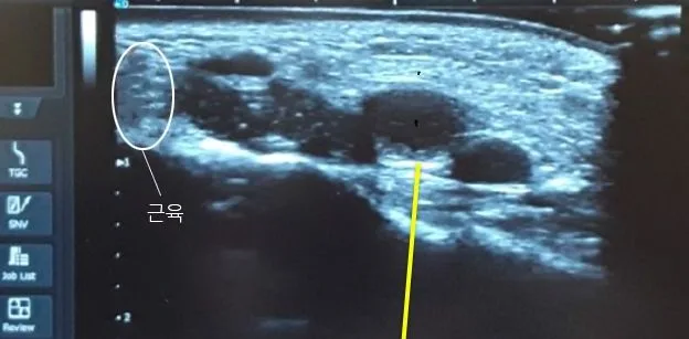
- Ultrasound shows the depth and distribution of material, while CT identifies hardened areas and changes in bone.
- How are they removed?
- A fine cannula, ultrasound equipment, endoscopy, or an incision is selected according to whether the material is soft or hard and how firmly it adheres to surrounding tissue.
- Can everything be removed?
- 100% removal is difficult because nerves and normal tissue must be preserved. Priority is given to reducing the areas causing pain, inflammation, and deformity.
How this differs from conventional methods
Conventional methods
When filler forms lumps or causes swelling, dissolving injections are often tried first. Steroid injections may be used to reduce inflammation, and material may also be expressed or aspirated through a needle or fine cannula.
Limitations
Dissolving injections work only on hyaluronic acid fillers. They do not dissolve fillers with other ingredients, such as Ellansé or Artecoll, grafted fat, or unapproved materials. Steroid injections reduce swelling and inflammation, but the causative material remains, so problems may recur over time.
Material that has hardened over time or become adherent to surrounding tissue is difficult to remove without seeing it. Material may remain, or normal tissue may be injured during removal.
Dr. Kang's approach
Ultrasound and CT are first used to identify the location, depth, and amount of material. The method is then selected according to its condition. Soft material with little adhesion is removed with a fine cannula; adherent material is separated using ultrasonic vibration. An endoscope provides a magnified view in the nerve-rich forehead, temples, and glabella, while hard lumps are removed under direct visualization through an incision.
The appearance after removal is planned in advance because the area may become hollow or the skin may sag.
Benefits and precautions
Removal under direct visualization allows problematic material to be selectively reduced while preserving nerves, blood vessels, and normal fat. Identifying its location beforehand helps avoid unnecessarily extensive dissection.
Complete removal is not the goal. Aggressive removal can cause depressions or nerve injury, so some material may be left in place or surgery may be staged. Recovery usually takes at least 1 week, and the under-eye area is monitored for at least 6 months.
Glossary
- Hyaluronidase
- An injectable medication that dissolves hyaluronic acid fillers. It does not work on fillers with other ingredients.
- Foreign-body granuloma
- A firm inflammatory lump that forms as the body surrounds foreign material.
- Adhesion
- A condition in which tissues that should be separate become stuck together.
- Calcification
- Calcium deposits in tissue that cause it to become hard like stone.
- Ultrasound examination
- An examination in which a probe placed on the skin displays the underlying tissues on a screen. It identifies the depth and extent of material.
Types of material
The approach depends on the material. Hyaluronidase dissolves only fillers composed of hyaluronic acid. It cannot break down semipermanent fillers made of collagen or calcium, which are removed surgically.
| Category | Examples | Characteristics |
|---|---|---|
| Illicit foreign materials | Candle wax, industrial paraffin, industrial silicone, petroleum jelly, oils, illicit collagen, illicit fillers | These materials are not approved. They are highly likely to adhere to tissue, and even without symptoms, the likelihood of chronic inflammation, skin necrosis, and granulomas increases with delay. |
| Permanent fillers | Aquamid, Royamid, Perform | Infection and skin necrosis can occur. If there is an inflammatory reaction, removal may follow a course of medication. |
| Collagen-stimulating fillers | Ellansé | A filler combining PCL and CMC. If more collagen forms than expected, it can harden together with surrounding cells. |
| PMMA fillers | Artecoll, Artefill | The ingredient is also used in artificial bone. Over time, it can form hard lumps that are very difficult to remove. Direct incisions or endoscopy are more suitable than a cannula. |
| Calcium fillers | Radiesse | They have the advantage of limited migration, but strong tissue adhesion makes removal difficult. |
| Hyaluronic acid fillers | When hyaluronidase does not dissolve them | Most can be dissolved by injection, but longstanding filler or advanced fibrosis may require surgical removal. |
| Grafted fat | Excessive graft retention, calcification, fibrosis, oil cysts | Symptoms may emerge over time, and weight gain can make the grafted area appear disproportionately enlarged. |
Fillers that UVOM Plastic Surgery lists as not dissolvable and candidates for surgical removal
Diagnosis
Preoperative ultrasound identifies the actual distribution and depth of foreign material. This helps reduce unnecessary dissection and injury to normal tissue while enabling precise removal of the foreign material.
3D CT shows the location and amount of calcification and cysts. A hard lump after fat grafting is calcified if it appears with bone density on CT. For forehead implants, CT assesses position, size, and bone erosion.
Removed tissue is sent for microscopic examination when necessary. This can identify inflammation, firm lumps, or hardened tissue around the foreign material.
Removal methods
No single method can remove every type of foreign material. Methods are selected according to firmness, adhesion, and location, and combined when necessary.
Removal through a fine tube (cannula)
If the material is not hard and has little adhesion to surrounding tissue, ultrasound identifies its precise location before a fine cannula is inserted to remove it without an incision.
Ultrasound-assisted removal
Ultrasonic vibration separates the material from surrounding tissue before removal. Excess grafted fat is selectively liquefied and removed evenly. The likelihood of an uneven skin surface is low.
Endoscopic removal
These areas contain many nerves, and motor nerves pass from the temples toward the forehead. Removal is performed under 20-fold magnification while preserving nerve tissue. Incisions within the scalp reduce visible scarring.
Removal through an incision
Hardened foreign material and granulomas are removed under direct visualization. Around the eyes and nose, removal is combined with related surgery to reduce incisions; for the lips and deep cheeks, incisions inside the mouth keep scars out of view.
Laser and similar methods achieve lower removal rates for fillers that cannot be dissolved. Surgery through an incision or with an endoscope allows direct assessment of the amount, helping remove a substantial quantity safely and reduce the risk of vascular and nerve injury.
Approach by area
Incisions use existing surgical scars or inconspicuous sites.
| Area | Approach |
|---|---|
| Around the eyes | An incision is made below the eyebrow or under the eye. Upper or lower blepharoplasty or under-eye fat repositioning may be combined when appropriate. Some materials can be removed through a small opening. |
| Nose | An open approach is used to remove material while protecting the structures. Cartilage grafting is used to prevent contracture that can lift the nasal tip as tissue contracts. |
| Lips | An incision inside the lip allows removal of adherent masses, nodules, and dispersed foreign material. |
| Forehead | An incision in the scalp above the hairline allows endoscopic removal. A cannula is used in some cases. |
| Cheeks, deep cheeks, and nasolabial folds | Foreign material in the subcutaneous fat layer is removed with a cannula; material in the deep cheek is removed through an incision inside the mouth. |
| Chin | Material is removed with a cannula or through a skin incision beneath the chin. Deeper material is approached through an incision inside the mouth. |
| When skin necrosis has progressed | Because surgery is performed on already damaged tissue, concurrent treatment with fat-derived regenerative cells (SVF) is needed. |
Under-eye foreign material removal
Under-eye foreign material removal at UVOM Plastic Surgery is performed exclusively by Medical Director Dr. Chang Min Kang.
Regarding under-eye foreign material removal, Medical Director Dr. Chang Min Kang explains, “Because the skin is thin, I believe a more thorough and clean removal than in other areas is needed for greater satisfaction with surgery.”
The under-eye skin is thinner than in other areas, making foreign material easier to see and feel. Numerous nerves and blood vessels lie within the muscle, and foreign material may be entangled with them. Surgery must account for the relationship between the foreign material and normal structures to prevent lower eyelid retraction (ectropion), which distorts the lower eyelid.
What feels like one lump from the outside may appear during surgery as multiple large and small capsules among the orbicularis oculi muscle fibers. Once encapsulation has developed, non-incisional removal is more likely to leave material behind. Residual material is more noticeable under the eyes than elsewhere, so removal of a substantial amount through an incision is preferable.
When needed, the approach used for lower blepharoplasty can remove foreign material while also correcting under-eye structures. Material spread widely between the skin and muscle may be difficult to remove completely in one operation, and some may remain. Excessive removal may cause hollowing or alter the lower eyelid shape. Patients often do not remember the product name of injections received years or decades ago; ultrasound may also be unable to identify the precise composition of old mixtures and fibrosis. In such cases, diagnosis and treatment proceed together while assessing the response to hyaluronidase. If pain, deformity, and inflammation persist, surgery and tissue examination are considered.
Removal methods
- Non-incisional approach When material is soft and not far from the under-eye area, it is selectively liquefied and removed through a small opening.
- Transconjunctival incision Suitable when skin laxity is limited and the material is not far from the eye.
- Skin incision Used when material is hard, skin laxity needs correction, or the location is farther from the under-eye area, such as the anterior cheek, cheekbone, or tear trough.
| Item | Under-eye foreign material removal | Grafted fat removal |
|---|---|---|
| Operating time | Within 90–120 minutes | Within 60 minutes |
| Anesthesia | Sedation | Sedation |
| Suture removal | After 5–7 days | After 7 days, if sutures are present |
| Follow-up treatment visits | 1–2 visits | 1–2 visits |
| Hospital admission | Not required | Not required |
| Recovery period | After approximately 1 week | After approximately 1 week |
These are UVOM Plastic Surgery's general guidelines and may vary with the patient's condition and surgical method.
Inflammation
Swelling, warmth, and pain under the eyes
Deformity
An uneven under-eye contour or an outward-turned lower eyelid
Visible foreign material
Injected material that shows through the skin or can be felt
Adverse reactions
Peeling or oozing skin, or palpable firm nodules
Skin necrosis
Vascular compression causes the treatment area to turn bluish or develop blisters and tissue death
Distinguishing it from under-eye fat
Appearance or touch alone cannot distinguish them. Procedure history, an in-person examination, and high-frequency ultrasound are used.
Removal of grafted fat
Complications of fat grafting can emerge over time. Grafting a large volume at once can cause sagging from the weight of the fat cells. With weight gain, fat cells in the grafted area may enlarge and make that area look disproportionately bulky.
| Symptom | Description |
|---|---|
| Excessive graft retention | Excessive grafting creates an unnatural or asymmetrical appearance. |
| Oil cysts | If the center of grafted fat tissue loses its blood supply and becomes necrotic, it can form a cyst. It feels firm like a nodule and contains oil. |
| Inflammation and infection | Excessive postoperative swelling persists, with warmth and edema. |
| Calcification | Calcium from the blood deposits in the tissue, making it hard and uneven. In severe cases, irritation of nearby nerves causes pain. |
| Fat necrosis | Fat that fails to survive can cause skin infection and tissue necrosis, which may discolor the skin surface. |
Extent of removal
Not all fat is removed indiscriminately; tissue near nerves and muscles must be considered. If it cannot be felt by hand, excessive removal is best avoided. Aggressive removal can injure tissues, including nerves.
Timing of further removal
Swelling and bruising increase for about 3 days after removal before subsiding. After an intermediate period of 6–8 weeks, swelling continues to decrease for up to 6 months. If further removal is still needed after this period, an additional procedure is possible.
Forehead implant removal
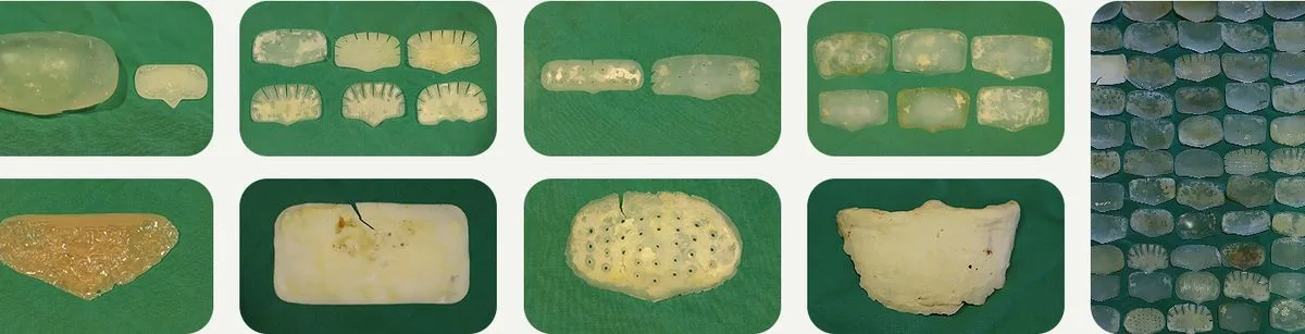
Forehead implants are often custom-made to fit an individual's forehead. Materials include silicone, Gore-Tex, bone cement, and AlloDerm, and even implants of the same material vary in size and shape. UVOM Plastic Surgery has used endoscopy to remove forehead implants since 1999.
Surgical principles
- Use the existing incision. The previous surgical site is used to avoid adding scars. If additional incisions are needed, 1–3 sites of approximately 2–3 cm are used.
- Remove bony projections. Bone can grow through small holes in silicone implants. Bony projections that could be felt after surgery are removed. A prominence of about 1 mm is cushioned by skin thickness and cannot be felt.
- Check twice. The removed implant's shape is checked immediately. If a piece is missing or torn, a second endoscopic check looks for retained fragments.
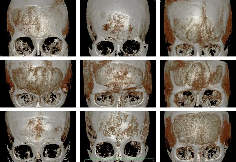
Preoperative CT identifies the implant's position and size and the locations where nerves emerge. Bone erosion can develop over time after implant surgery, so potential forehead hollowing after removal is anticipated.
When removal is considered
- Blood pooling or a hematoma
- Persistent headache or a foreign-body sensation
- Implant displacement causing asymmetry
- An artificial appearance or a visible implant edge
- When considering a forehead lift
Procedures considered together
- Endoscopic forehead lift Lifting with an implant in place increases the likelihood of complications from displacement. Combining the lift with removal uses the existing dissection space and reduces unnecessary dissection.
- Forehead fat grafting Removal can leave volume loss or irregular contours caused by bone erosion. Fat grafting is recommended 6 months after surgery.
Witch's chin (chin ptosis)
Soft tissues including muscle, fibrous tissue, and fat cover the chin bone and shape the chin. When these tissues sag below the chin, it looks long and drooping, often more noticeably when smiling. This appearance is commonly called a witch's chin.
Chin surgery usually involves an incision inside the mouth and detachment of muscles from the chin bone before cutting the bone or placing an implant. If muscle and skin elasticity are not considered or muscle closure is inaccurate, remaining muscle and scar tissue can sag, causing chin ptosis, a double chin, or a witch's chin. It may also occur after filler or other foreign material injections or fat grafting, or when soft tissue is naturally positioned lower.
| Method | Suitable situations |
|---|---|
| Chin osteotomy | A long or wide chin bone is shortened or narrowed; a protruding chin is moved backward, and a recessed chin is moved forward. |
| Chin augmentation | A short chin bone is augmented with an autologous dermal graft, an artificial dermal graft, or an implant. |
| Chin lift (witch's chin correction) | If bone surgery is burdensome or sagging is the main concern, an incision beneath the chin is used to reduce and elevate drooping tissue. |
Steps of a chin lift
- Design based on length proportions Using a ratio of 1 for the distance from beneath the nose to the lip line and 1.618 from the lip line to the chin as a reference, the design connects the jawline and chin in profile.
- Concealed incision An incision on the underside of the chin is kept out of view from the front and does not extend beyond the chin.
- Careful dissection Dissection preserves an appropriate skin thickness to avoid unevenness.
- Removal of excess tissue Excess drooping tissue below the chin is excised.
- Closure Tissue and skin are drawn up, secured, and sutured to blend smoothly with the surrounding jawline.
When this can occur
- Naturally low-hanging soft tissue
- Loss of elasticity and loosening after chin surgery
- Soft-tissue stretching from excessive procedures or implants
- Deformity caused by granulomas from filler or foreign material
- A chin that elongates when smiling
- A drooping, dumbbell-shaped chin with protrusions on both sides
Limitations and recovery
Can surgery remove 100% of the material?
Even with surgery, 100% removal is difficult because normal tissue must be preserved. However, adherent foreign material can be removed under endoscopic or direct visualization, and hardened granuloma masses can also be removed while protecting nerves, blood vessels, and muscles.
Can I recover immediately after surgery?
Swelling increases for 48–72 hours after surgery, then gradually subsides. Although recovery varies, everyday activities can generally resume after an average of 7 days. Severe bruising may require 2 weeks or longer.
Is swelling worse under the eyes?
Swelling peaks on day 2. Applying an ice pack over clean gauze can help. Scar tissue may make the skin uneven, so tissue softening can take longer than after other cosmetic procedures. Follow-up for at least 6 months is recommended while swelling and bruising settle.
Can semipermanent fillers only be removed surgically?
Surgery is not always the only option. A non-incisional method may be effective in 1 session, but a 2nd or 3rd may be needed. Because problematic filler is best removed promptly, the method is selected according to the condition.
Why is an endoscope used for filler removal?
Endoscopy is used for the forehead, temples, and glabella. These areas contain many nerves, and motor nerves pass from the temples toward the forehead. Injury along a nerve's course can cause paralysis, so endoscopy is used to preserve nerve tissue during removal.
Can I keep receiving dissolving injections if the material is unknown?
If lumps and a foreign-body sensation remain after an adequate course of dissolving treatment guided by a clinician's diagnosis, surgical removal may be worth considering. If residual foreign material causes recurrent inflammation and deformity, removal under direct visualization of the material and altered tissue can be considered.
Blood collected after forehead implant surgery and was drained with a syringe. Does the implant need to be removed now?
If pressure or headache resolves, observation may be possible for a while. However, removal is recommended because blood pooling often recurs. It occurs when bone erosion creates a space between the implant and forehead bone, and this space may enlarge further.
Can I apply hot compresses after surgery?
This is not recommended. Reduced sensation at the surgical site creates a risk of burns. After cold compresses for 2–3 days, a warm half-body bath is recommended to help circulation and lymphatic drainage.
Is it best to remove all facial foreign material at once?
No. Trying to remove all material spread across several layers or mixed with normal fat, muscle, and nerves may increase the risk of depressions, functional changes, and nerve injury. Prioritizing areas causing pain, inflammation, and deformity, leaving some material, or staging surgery may be safer.
What do ultrasound and CT each assess?
Ultrasound is useful for viewing the location and depth of material in soft tissue, nodules and fluid-related changes, and relationships with surrounding tissue in real time. CT helps assess bone position, calcification, previous facial contouring surgery, and extensive deformity. The examinations needed depend on symptoms and surgical history.
Can dissolving injections be tried first?
Hyaluronidase may be considered first when hyaluronic acid filler is confirmed or strongly suspected. However, old mixtures, fat-graft lumps, fibrosis, and granulomas may respond only to a limited extent. Before repeating injections, it is important to reassess the tissue layer containing the material and the inflammatory condition. If hyaluronic acid is confirmed and dissolving injections can resolve the problem, surgery is not rushed.
Why can the face sag more after foreign material removal?
Removing material that has filled facial volume for a long time can leave space and excess skin. If fat and the SMAS have already shifted downward, sagging may become more evident after removal. Remaining volume and supporting layers are therefore assessed in advance.
Can foreign material removal and a facelift be performed on the same day?
Sometimes, but this is not appropriate for every patient. Active inflammation, infection risk, extent of material, skin blood supply, and anticipated dissection are assessed. Combined surgery may be considered if substantial sagging is expected after removal of localized material and inflammation is stable. If risk is high, surgery is staged.
Care after removal
Depending on the area, a forehead lift, facelift, or thread lift may be considered for skin and muscle laxity caused by foreign material. Fat grafting may improve hollow or uneven areas after removal. Ultrasound lifting is recommended 3 months after surgery. Recommendations vary by age and area.

Surgery and consultation
Consultations are provided by Medical Director Dr. Chang Min Kang, a plastic surgery specialist. About the doctor
View each study's population, methods, results, and limitations
Supporting evidence
- Evidence card: facial foreign material removal
- 2020 presentation on endoscopic foreign material removal cases · 2017 presentation on foreign-body granuloma removal
- Doctor's Columns by Medical Director Dr. Chang Min Kang: “How can under-eye foreign material be distinguished from under-eye fat?” “How can an old facial injection be assessed when its ingredients are unknown?” “Can under-eye foreign material removal and lower blepharoplasty be performed together?”
- UVOM Plastic Surgery academic presentations: 2020 PRS KOREA, “Endoscopic removal of foreign material that could not be removed by non-incisional methods” (Ik Gyun Choi, Dong Hyun Yoon, Jung Su Lee); 2017, “Removal of an asymptomatic foreign-body granuloma after cutaneous HA injection”
Recovery course
| Timing | Typical course |
|---|---|
| 2–3 days after surgery | Swelling is at its worst. Under-eye swelling peaks on day 2, and cold compresses can help. |
| 5–7 days after surgery | Sutures are removed if an under-eye incision was made. Everyday activities can generally resume after an average of 7 days. |
| 2 weeks after surgery | Severe bruising may take this long to settle. |
| 6–8 weeks after surgery | An intermediate phase during which swelling steadily decreases. |
| 3 months after surgery | Treatments such as ultrasound lifting can begin. |
| 6 months after surgery | Most swelling has subsided, and the need for further removal is assessed. The under-eye area is monitored for at least 6 months. |
This may vary with the individual's condition and the extent of surgery.
Questions answered by Dr. Kang
Questions answered directly by Medical Director Dr. Chang Min Kang in the Doctor's Columns.
Can filler injected long ago still remain?
Yes, it can.
Hyaluronic acid fillers generally break down over time, but may persist longer than expected depending on the product's properties, amount and location injected, repeated treatments, and the individual's tissue condition.
However, not all fillers break down in the same way. If the composition is unknown or a non-hyaluronic acid material was used, persistence and treatment may differ.
A long interval since injection therefore does not establish that all the filler has disappeared.
Answered by Medical Director Dr. Chang Min Kang · Could firmness under the eyes be caused by old filler?
Does firmness under the eyes always mean filler?
No.
The remaining filler itself may be palpable, or the cause may be filler clumping, surrounding adhesions or fibrosis, or an inflammatory reaction.
Skin or subcutaneous cysts, scar tissue, and other causes unrelated to filler may produce similar symptoms, so an examination is needed to distinguish them.
Answered by Medical Director Dr. Chang Min Kang · Could firmness under the eyes be caused by old filler?
Does all old under-eye filler need to be removed?
The presence of residual filler does not always require removal.
If there are no specific symptoms or concerns about appearance, observation may be appropriate depending on the condition.
However, persistent firm lumps, recurrent swelling, pain, a foreign-body sensation, asymmetry, or uneven contours warrant assessment of the filler location and surrounding tissue before deciding whether treatment is needed.
With old filler, the current under-eye condition matters.
Under-eye firmness may reflect residual old filler, but filler clumping, surrounding adhesions and fibrosis, nodules, and inflammatory reactions may also contribute.
Especially after treatment long ago or repeated filler injections, the original treatment information alone may not establish the current condition.
Because under-eye skin and soft tissues are thin and move repeatedly with facial expressions, even small changes can readily affect appearance and feel.
If firm lumps, recurrent swelling, pain, a foreign-body sensation, or unevenness persist, assessment of possible residual filler, its location, and surrounding tissue is recommended before discussing suitable treatment.
If lumps, firmness, recurrent swelling, or a foreign-body sensation persist after old under-eye filler injections, you can arrange a consultation with UVOM Plastic Surgery at 02-545-3700 or via KakaoTalk at “UVOM Plastic Surgery.”
※ Diagnosis and treatment may vary with filler composition and location, surrounding tissue condition, and individual symptoms.
Answered by Medical Director Dr. Chang Min Kang · Could firmness under the eyes be caused by old filler?
Can under-eye filler remain for at least 10 years?
It is possible. An MRI study of the midface reported cases in which hyaluronic acid filler was visible for up to 15 years.
However, the study did not focus exclusively on the under-eye area and included few participants, so it cannot be generalized to mean all under-eye filler persists for at least 10 years.
Answered by Medical Director Dr. Chang Min Kang · Can filler injected under the eyes long ago still remain?
Can you tell by touch whether filler remains?
Filler near the skin or concentrated in one area may be palpable, but material spread widely in deeper layers is difficult to confirm by touch alone.
High-frequency ultrasound may help with diagnosis when necessary.
Answered by Medical Director Dr. Chang Min Kang · Can filler injected under the eyes long ago still remain?
Can ultrasound identify the exact type of filler?
Ultrasound helps identify the location and distribution of suspected filler or foreign material, but cannot definitively establish the product name or composition of every filler.
Past treatment records and examination findings must also be considered.
Answered by Medical Director Dr. Chang Min Kang · Can filler injected under the eyes long ago still remain?
Can all old under-eye filler be dissolved in one session?
This depends on filler type, amount, location, distribution, and repeated treatments. Old filler or filler distributed across several layers may require staged treatment.
Answered by Medical Director Dr. Chang Min Kang · Can filler injected under the eyes long ago still remain?
Will dissolving filler immediately restore my original under-eye appearance?
Not always. As filler volume decreases, the original tear trough or protruding under-eye fat may become visible again. If surrounding swelling or inflammatory nodules remain, the appearance may not recover immediately.
Answered by Medical Director Dr. Chang Min Kang · Can filler injected under the eyes long ago still remain?
Does old residual filler always need removal?
Not necessarily if there are no symptoms or cosmetic concerns. Treatment decisions should be based on problems such as edema, nodules, inflammation, pain, and asymmetry rather than the presence of filler alone.
Answered by Medical Director Dr. Chang Min Kang · Can filler injected under the eyes long ago still remain?
Does firmness under the eyes always indicate foreign material?
No. Firm nodules can have various causes, including fibrosis around injected material, a foreign-body reaction, fat necrosis after fat grafting, oil cysts, calcification, and scar tissue.
Touch alone cannot establish a diagnosis. Procedure history, examination, and imaging when needed are required.
Answered by Medical Director Dr. Chang Min Kang · How can under-eye foreign material be distinguished from under-eye fat?
Can filler injected long ago still remain under the eyes?
Yes. Persistence varies with filler composition, injected volume and depth, and individual tissue reactions.
Even if treatment was long ago, tell your clinician about previous filler injections if you have under-eye swelling or bulging.
Answered by Medical Director Dr. Chang Min Kang · How can under-eye foreign material be distinguished from under-eye fat?
Can ultrasound detect under-eye filler?
High-frequency ultrasound is useful for assessing the possible presence, location, depth, and distribution of injected materials such as filler, as well as surrounding tissue.
However, imaging alone cannot always establish the exact product or composition, so past treatment records and examination findings are assessed together.
Answered by Medical Director Dr. Chang Min Kang · How can under-eye foreign material be distinguished from under-eye fat?
Can under-eye fat and filler be present at the same time?
Yes. Filler may have been injected over existing protruding orbital fat, or filler-related swelling and skin laxity may coexist.
Their respective locations and effects must be distinguished rather than assuming a single cause.
Answered by Medical Director Dr. Chang Min Kang · How can under-eye foreign material be distinguished from under-eye fat?
Must all under-eye foreign material be removed?
Not every injected material requires removal. Symptoms, deformity, inflammation, and the type and location of material must be considered.
When material is severely adherent to normal tissue, complete removal may increase tissue injury risk. Whether and how much to remove must therefore be decided individually.
Answered by Medical Director Dr. Chang Min Kang · How can under-eye foreign material be distinguished from under-eye fat?
Can dissolving injections remove all under-eye filler?
Hyaluronidase is used to break down hyaluronic acid fillers.
However, it does not eliminate all non-hyaluronic acid fillers, nonabsorbable materials, post-fat-grafting nodules, or established fibrotic tissue. The material used and current tissue condition must be assessed first.
Under-eye fat and foreign material cannot be distinguished by appearance or touch alone.
Three points are important for accurate differentiation.
- Review previous filler, fat-grafting, and other injection procedures.
- Examine the location of under-eye bulging and eyelid structures in person.
- When needed, use imaging such as high-frequency ultrasound to identify the material's location and surrounding tissue condition.
Orbital fat, injected materials such as filler, post-fat-grafting changes, fibrosis, swelling, and skin laxity may coexist under the eyes.
Before selecting treatment, the priority is therefore to identify which tissue layer the bulging arises from and which cause contributes most.
Accurate diagnosis helps reduce unnecessary filler dissolution, excessive fat removal, and repeated procedures, and enables a treatment plan suited to the current under-eye condition.
For questions about distinguishing under-eye fat from foreign material, please call 02-545-3700 or contact “UVOM Plastic Surgery” on KakaoTalk.
For more information on under-eye foreign material, click a title below ▼
Can under-eye foreign material removal and lower blepharoplasty be performed together?
Why do my under eyes keep looking swollen after filler injections?
Answered by Medical Director Dr. Chang Min Kang · How can under-eye foreign material be distinguished from under-eye fat?
Does under-eye filler prevent me from having lower blepharoplasty?
No. Depending on the location of filler or foreign material and the tissue condition, procedures may be performed together on the same day or in stages.
Answered by Medical Director Dr. Chang Min Kang · Can under-eye foreign material removal and lower blepharoplasty be performed together?
Can removing foreign material first change the lower blepharoplasty plan?
Yes. Removing the volume occupied by foreign material changes the skin and fat condition. The amount of skin to excise may be reassessed after removal.
Answered by Medical Director Dr. Chang Min Kang · Can under-eye foreign material removal and lower blepharoplasty be performed together?
Do dissolvable fillers also require surgical removal?
Dissolving hyaluronic acid filler with hyaluronidase injections is considered first. However, surgical removal may be needed in some longstanding cases or when fibrosis has developed.
Answered by Medical Director Dr. Chang Min Kang · Can under-eye foreign material removal and lower blepharoplasty be performed together?
Does combined surgery take longer to recover from?
Initial swelling may be greater than after lower blepharoplasty alone. However, the overall recovery period is often shorter than with two separate operations.
Answered by Medical Director Dr. Chang Min Kang · Can under-eye foreign material removal and lower blepharoplasty be performed together?
Will there be more scarring?
If the lower blepharoplasty incision can be used, an additional incision may not be needed. However, a separate approach may be necessary if foreign material lies beyond the incision's reach.
Answered by Medical Director Dr. Chang Min Kang · Can under-eye foreign material removal and lower blepharoplasty be performed together?
Can a consultation alone determine whether the procedures can be combined?
A consultation alone is generally insufficient. Surgery is usually planned after ultrasound assesses the material's location and extent and the presence of inflammation.
Under-eye foreign material removal and lower blepharoplasty can sometimes be performed together. However, the same approach does not apply to every patient.
Before deciding whether surgery can be combined, the material's location and the tissue condition must be assessed.
Depending on ultrasound findings, same-day surgery may be possible, or it may be more appropriate to remove the material first and perform lower blepharoplasty after tissue recovery.
The material type, location, and tissue response differ between patients. Planning the extent and sequence of surgery based on examination findings helps achieve a safe outcome.
If you are considering both under-eye foreign material removal and lower blepharoplasty, please call 02-545-3700 or contact “UVOM Plastic Surgery” on KakaoTalk.
For more columns on under-eye foreign material removal, click a title below ▼
Why do my under eyes keep looking swollen after filler injections?
Answered by Medical Director Dr. Chang Min Kang · Can under-eye foreign material removal and lower blepharoplasty be performed together?
Will swelling from under-eye filler improve on its own over time?
Normal swelling immediately after treatment usually improves within 1–2 weeks. If it persists beyond this period, the filler location and amount, as well as structural causes in the under-eye area, should be assessed.
Answered by Medical Director Dr. Chang Min Kang · Why do my under eyes keep looking swollen after filler injections?
Can swelling develop years after under-eye filler injections?
Yes. Hyaluronic acid filler can remain longer than expected. Over time, its attraction of water or changes in position may make swelling noticeable later. Clinical examination and ultrasound are recommended to establish the cause.
Answered by Medical Director Dr. Chang Min Kang · Why do my under eyes keep looking swollen after filler injections?
Can salty food or alcohol make under-eye filler look more swollen?
Yes. Because hyaluronic acid filler attracts water, sodium intake, alcohol, or changes in physical condition can temporarily make swelling more noticeable.
Answered by Medical Director Dr. Chang Min Kang · Why do my under eyes keep looking swollen after filler injections?